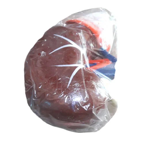
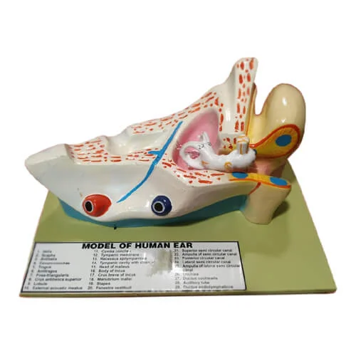
Human Duodenum Model on Board – Anatomical Digestive System Teaching Model
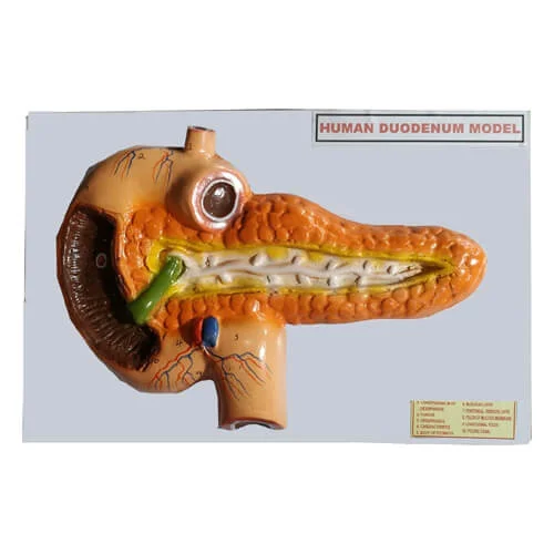
The Human Duodenum Model on Board is an educational relief teaching model engineered to demonstrate the microanatomy, ductal junctions, and retroperitoneal relationships of the human duodenum—the vital proximal first segment of the small intestine. Cast in life-size relief from high-density, unbreakable medical PVC polymer and mounted securely on a durable demonstration board, this model accurately represents the upper gastrointestinal transition zone where gastric chyme mixes with biliary and pancreatic digestive secretions. It serves as an essential teaching benchmark across secondary school biology classrooms (SSC/HSC), university physiology departments, nursing academies, and medical colleges throughout Bangladesh.
Understanding duodenal histology, peptic ulcer formation zones, and pancreatobiliary ductal drainage pathways can be challenging when relying solely on two-dimensional textbook sketches. This relief board model resolves that learning barrier by providing an open, interconnected dissection view of the C-shaped duodenal loop (divided into superior, descending, inferior, and ascending parts). The stomach's pyloric region is shown opening through the pyloric sphincter into the duodenal bulb (cap), while the head of the pancreas nests snugly within the inner curvature of the loop.
A prominent anatomical highlight is the internal coronal cutaway of the duodenal wall, which exposes circular mucosal folds (plicae circulares), villi, and the critical major duodenal papilla where the common bile duct and main pancreatic duct unite. The model also traces the tortuous course of the superior mesenteric vessels and pancreaticoduodenal arterial arches. Each critical landmark is marked with numbered identification callouts linked to a printed reference index card. Victor Scientific Co. supplies high-grade anatomical and surgical teaching models nationwide with guaranteed quality and secure packaging.
✹ Key Features
C-Shaped Duodenal Loop Architecture
Presents realistic adult anatomical proportions showing all four distinct anatomical parts of the duodenum surrounding the pancreatic head.
Internal Mucosal Plicae Circulares Detailing
Coronal cutaway of the intestinal wall reveals internal circular folds (plicae circulares) and the major duodenal papilla (ampulla of Vater).
Hepatopancreatic Ductal Convergence
Clearly depicts the junction where the common bile duct from the gallbladder and the main pancreatic duct unite before entering the duodenal lumen.
Integrated Regional Vasculature
Shows adjacent regional blood vessels including the gastroduodenal artery, superior mesenteric artery, and portal venous branches.
Durable, Medical-Grade PVC Build
Cast from high-density, washable medical PVC plastic mounted on a finished wooden or heavy-duty polymer baseboard that resists tropical humidity.
Vibrant Anatomical Color Coding
Uses fade-resistant, non-toxic colors that clearly contrast the duodenal wall (pink/buff), pancreas (yellowish-tan), bile ducts (green), and blood vessels (red/blue).
Numbered Demonstration Key Card
Features clear numerical callouts stamped beside each anatomical structure corresponding to an included laminated reference legend for spot tests.
Wall-Mountable and Benchtop-Ready
Designed with a balanced footprint suitable for demonstration easels or secure wall hanging in school science, nursing, and medical laboratories.
✹ Applications
- Medical college MBBS anatomy practicals, gastroenterology lectures, and student revision
- Nursing institutes, paramedic academies, and allied healthcare foundation training
- Gastroenterology and general surgery outpatient departments (OPD) for patient education
- Higher Secondary (HSC) and university undergraduate biology and human physiology classrooms
- Interactive STEM science exhibitions, medical fairs, and student viva-voce assessments
✹ Why Choose Human Duodenum Model on Board?
- Relief board mounting provides an unobstructed, complete structural overview without loose, easily misplaced pieces.
- High-contrast paint finish makes fine ampullary openings and mucosal folds visible across large classrooms.
- Eliminates the biohazard handling, chemical odors, and tissue decay of preserved cadaveric prosections.
- Solid PVC construction withstands continuous handling by multiple student cohorts.
- Authentic institutional procurement and dependable stock availability backed by Victor Scientific Co.
✹ Benefits
- Accelerates student comprehension of enzymatic digestion, duodenal ulcer sites, and biliary drainage.
- Enhances patient communication regarding ERCP procedures, peptic ulcer disease, and duodenal obstruction.
- Delivers a reusable, permanent teaching asset that serves multiple academic cohorts without recurring costs.
- Speeds up student assessment during laboratory spot practicals and viva-voce examinations.
✹ About Victor Scientific Co.
Victor Scientific Co. provides quality laboratory equipment, measuring instruments and scientific tools in Bangladesh. We supply reliable products for educational institutions, industries, laboratories and professional users.
 Health And Safety
 Laboratory Equipment
 Educational Equipment
 Lab Chemicals & Reagents
 Meters & Testers
 Weight Scale & Balance
 Garments & Textile Testing
 Electrical & Electronics
 Civil & Construction
 Industrial Safety Equipment