Animal Mitosis Model on Board – Biology Teaching Aid
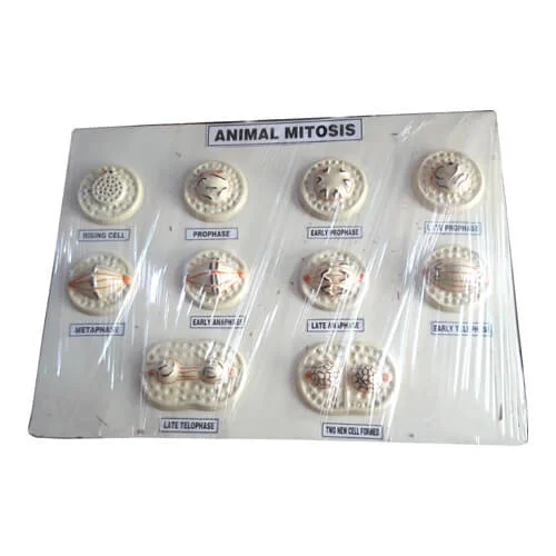
The Animal Mitosis Model on Board is a three-dimensional biological relief display designed to illustrate the mitotic cell cycle and somatic cell division in eukaryotic animal cells. Cast from high-density, unbreakable PVC plastic and mounted on a durable display board, this model magnifies cellular and nuclear transformations thousands of times. It serves as an essential teaching benchmark across secondary school biology classrooms (SSC/HSC), university zoology and biotechnology departments, and medical colleges throughout Bangladesh.
Understanding chromosome condensation, spindle apparatus formation, and equatorial alignment can be challenging for students when relying solely on flat textbook diagrams or fleeting glimpses through compound microscopes. This relief board model resolves that learning hurdle by presenting a clear step-by-step physical progression of karyokinesis and cytokinesis. The sequence accurately displays key stages: Interphase (nuclear envelope intact with chromatin), Early and Late Prophase (centriole duplication, asters forming, and chromosome condensation), Metaphase (chromosomes aligned along the equatorial plate with kinetochore spindle fibers), Anaphase (sister chromatids separating to opposite poles), Telophase (nuclear reformation), and Cytokinesis (formation of the contractile ring and cleavage furrow yielding two identical diploid daughter cells).
Each mitotic phase is sculpted in raised relief and finished with non-toxic, chip-resistant pigments that highlight chromosomes, centromeres, spindle fibers, centrioles, and asters. The board comes complete with numbered identification indices and a printed reference key card, making it ideal for classroom lecture demonstrations, group study, and practical viva-voce tests. Victor Scientific Co. supplies precision-crafted cytological models nationwide with guaranteed quality and secure packaging.
✹ Key Features
Complete Mitotic Sequence Representation
Displays 8 to 10 progressive relief phases covering Interphase, Prophase, Metaphase, Anaphase, Telophase, and daughter cell Cytokinesis.
High-Magnification 3D Anatomical Detailing
Magnifies microscopic cellular dynamics, clearly depicting chromosome condensation, centromere splitting, and mitotic spindle fibers.
Animal Cell-Specific Cytokinesis
Accurately demonstrates astral ray formation and the inward pinching of the plasma membrane (cleavage furrow), distinguishing animal mitosis from plant cell plate formation.
Durable, Impact-Resistant Build
Fabricated from medical-grade PVC plastic mounted on a finished wooden or heavy-duty polymer baseboard that resists cracking, chipping, and tropical humidity.
Vibrant Multi-Color Painted Finish
Uses fade-resistant, non-toxic colors that clearly distinguish chromosomes (red/blue), spindle fibers (white/yellow), and cytoplasm under standard laboratory lighting.
Numbered Demonstration Key Card
Features clear numerical indices stamped beside each cellular stage corresponding to an included laminated identification legend for exams and spot tests.
Wall-Mountable & Benchtop-Ready
Designed with a balanced footprint suitable for demonstration easels or secure wall hanging in school science and biology laboratories.
✹ Applications
- High school (SSC) and higher secondary (HSC) biology practical classes and board curriculum demonstrations
- University undergraduate departments of Zoology, Botany, Biochemistry, and Biotechnology
- Medical college basic science, histology, and pathology lecture halls
- Nursing and allied healthcare training institutes
- Interactive STEM science fairs, biology exhibitions, and teacher training workshops
- Laboratory spot practical examinations and student viva-voce assessments
✹ Why Choose Animal Mitosis Model on Board?
- Converts complex, sub-microscopic cell division cycles into an intuitive, physical learning tool.
- High-contrast colors make delicate chromosome movements visible across large classrooms.
- Replaces fragile, difficult-to-prepare microscopic onion root tip or animal tissue slides for introductory lessons.
- Ready for immediate classroom display with zero assembly or calibration required.
- Authentic institutional procurement and reliable tender support provided by Victor Scientific Co.
✹ Benefits
- Accelerates student comprehension of chromosome behavior, diploidy, and genetic constancy.
- Speeds up student assessment during laboratory spot practicals and viva-voce tests.
- Reusable, permanent teaching asset that serves multiple academic cohorts without wear or fading.
- Enhances visual engagement during interactive lectures and teacher training workshops.
✹ About Victor Scientific Co.
Victor Scientific Co. provides quality laboratory equipment, measuring instruments and scientific tools in Bangladesh. We supply reliable products for educational institutions, industries, laboratories and professional users.
 Health And Safety
 Laboratory Equipment
 Educational Equipment
 Lab Chemicals & Reagents
 Meters & Testers
 Weight Scale & Balance
 Garments & Textile Testing
 Electrical & Electronics
 Civil & Construction
 Industrial Safety Equipment