Dicot Stem Model on Board – Educational Plant Anatomy Teaching Model
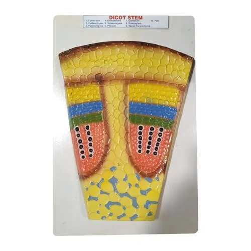
The Dicot Stem Model on Board is an educational botanical teaching aid engineered to demonstrate the internal histological organization, cellular differentiation, and primary tissue zones of a typical dicotyledonous stem (Helianthus annuus / sunflower stem). Cast in high relief from unbreakable, medical-grade PVC plastic or reinforced fiberglass and mounted securely on a finished display board, this model magnifies transverse section (T.S.) microscopic anatomy hundreds of times. It serves as an essential teaching benchmark across secondary school biology classrooms (SSC/HSC), college botany departments, agricultural universities, and plant physiology laboratories throughout Bangladesh.
Mastering plant histology, primary vascular arrangement, and the structural foundation for secondary growth is often difficult for students when relying solely on flat textbook drawings or delicate, hand-cut glass microscope slides. In dicot stems, tissue differentiation is well-defined into concentric zones, unlike monocot stems where vascular bundles remain scattered. This physical relief model resolves that learning barrier by providing an enlarged wedge sector and cross-sectional plan that exposes every cellular layer with anatomical fidelity.
The outer boundary features a single-layered epidermis with cuticular coating and characteristic multicellular uniseriate trichomes (epidermal hairs). Directly beneath lies the differentiated cortex, divided into an outer mechanical collenchyma hypodermis, chlorenchymatous/parenchymatous general cortex, and a wave-like single-layered endodermis (starch sheath). Inside the stele, the model clearly showcases the pericycle with sclerenchymatous patches forming hard bast caps over the vascular bundles. The open collateral vascular bundles are neatly arranged in a broken ring around a wide central pith, separated by prominent radial medullary rays. Each bundle clearly distinguishes external phloem, a multi-layered meristematic vascular cambium strip capable of secondary growth, and endarch xylem with large pitted metaxylem and interior spiral protoxylem vessels.
The board includes numbered identification indices paired with an attached laminated reference key card, making it ideal for classroom lectures, group study, and practical viva-voce assessments. Victor Scientific Co. supplies precision-crafted botanical and anatomical teaching models nationwide with guaranteed quality and secure packaging.
✹ Key Features
Magnified Transverse Section (T.S.) Relief
Displays an enlarged wedge sector and cross-sectional plan of a dicot stem, showing microscopic cellular architecture with dimensional clarity.
Concentric Ring Vascular Bundle Layout
Accurately demonstrates the eustelic organization of open collateral vascular bundles arranged in a neat circular ring around the central pith.
Active Vascular Cambium Layer
Clearly highlights the meristematic fascicular cambium strip situated between xylem and phloem, demonstrating the site of secondary vascular growth.
Detailed Multi-Layered Cortex
Differentiates the cuticularized epidermis with uniseriate trichomes, collenchyma hypodermis, parenchymatous cortex, and starch-rich endodermis.
Pericycle & Sclerenchymatous Hard Bast
Depicts semilunar patches of sclerenchyma cells capping the phloem, showcasing structural mechanical support tissues.
Durable, Impact-Resistant Construction
Fabricated from high-density, washable PVC plastic or fiberglass mounted on a finished wooden or heavy-duty polymer baseboard that resists tropical humidity.
Vibrant Botanical Color Coding
Finished with non-toxic, fade-resistant pigments that clearly contrast xylem (red/pink), phloem (blue/green), cambium (yellow/light green), and ground tissues.
Numbered Demonstration Key Card
Features clear numerical callouts stamped beside each anatomical tissue zone corresponding to an included laminated reference chart for practical exams.
Wall-Mountable and Benchtop-Ready
Designed with a balanced footprint suitable for demonstration easels or secure wall hanging in school botany and agricultural science laboratories.
✹ Applications
- Secondary School Certificate (SSC) and Higher Secondary (HSC) biology practical classes and board curriculum exams
- University undergraduate departments of Botany, Plant Breeding, Agronomy, and Forestry
- Agricultural universities and crop science research institutes for basic plant anatomy training
- Teacher training colleges, interactive STEM workshops, and science exhibition centers
- Agricultural extension training centers demonstrating dicot crop morphology (sunflower, cotton, pulses)
- Laboratory spot practical examinations and student viva-voce assessments
✹ Why Choose Dicot Stem Model on Board?
- Converts complex, sub-microscopic plant anatomy into an intuitive, tactile physical learning tool.
- High-contrast color mapping makes fine tissue layers visible across large lecture halls.
- Eliminates the preparation time, slide breakages, and staining errors associated with live plant microtome sectioning.
- Solid PVC construction withstands continuous handling by multiple student cohorts.
- Authentic institutional procurement and dependable stock availability backed by Victor Scientific Co.
✹ Benefits
- Accelerates student comprehension of dicot vs. monocot anatomical differences and secondary thickening.
- Speeds up student assessment during laboratory spot identification practicals.
- Delivers a reusable, permanent teaching asset that serves multiple academic cohorts without recurring costs.
- Enhances visual engagement during interactive lectures and teacher training workshops.
✹ About Victor Scientific Co.
Victor Scientific Co. provides quality laboratory equipment, measuring instruments and scientific tools in Bangladesh. We supply reliable products for educational institutions, industries, laboratories and professional users.
 Health And Safety
 Laboratory Equipment
 Educational Equipment
 Lab Chemicals & Reagents
 Meters & Testers
 Weight Scale & Balance
 Garments & Textile Testing
 Electrical & Electronics
 Civil & Construction
 Industrial Safety Equipment